
A new method to
Maximize the information extracted from flow-MRI data, using prior knowledge that the imaged object is a flow within a vessel network.
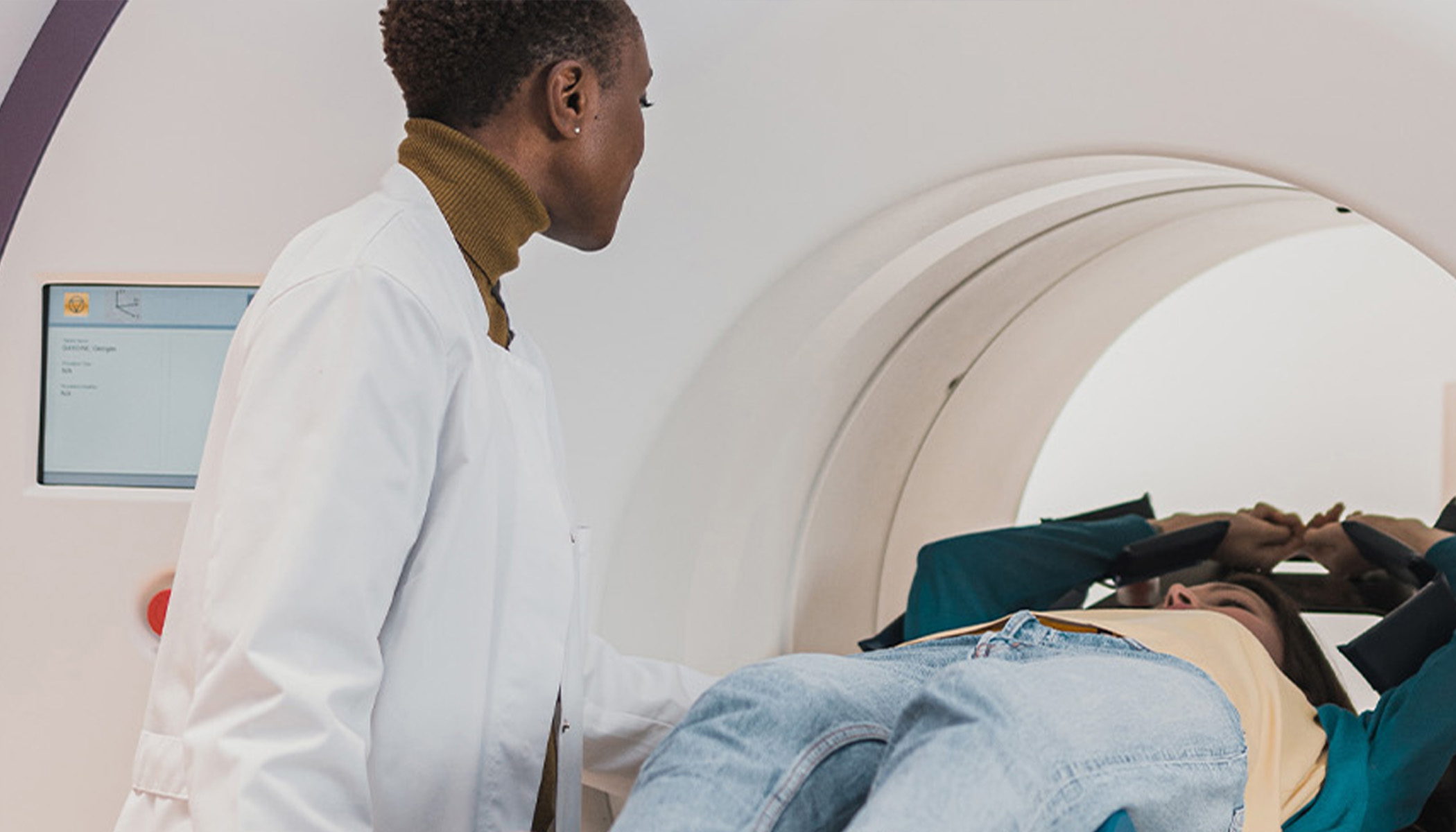
Flow-MRI allows clinicians to visualise flows in the body in 4D (3 spatial and 1 time dimension) without using ionising radiation. Currently, scans take tens of minutes and require the patient to remain still so that measurement noise can be reduced by averaging many images.
Even then, processed images remain noisy, particularly at the vessel boundaries. Accurate information is particularly valuable there because vessel wall shear stress is thought to be a major contributor to cardiovascular disease. Further, the measurement noise increases as the spatial resolution becomes finer, which means that flow-MRI is currently only used for large vessels. These factors have limited the clinical adoption of flow-MRI, despite its clear potential advantages in healthcare. Our technology addresses all of these limitations, greatly extending the potential clinical applications of flow-MRI.
Technology overview
Our Bayesian approach assimilates the flow-MRI data directly into a CFD simulation, using prior knowledge that the 3D image contains fluid flow through a vessel network. Unlike all other methods, it assimilates the vessel boundaries and the fluid velocity simultaneously, using flow information to refine the boundary location and boundary information to constrain the flow. This converges to the digital twin that is as close as possible to the patient’s flow-MRI scan, while also satisfying the equations of fluid flow. This reduces scan times by 10 to 100 times, converts measurement noise into quantified uncertainty, and outputs accurate wall shear stresses and pressure drops at no extra cost. Given that the capital cost of MRI infrastructure is high but the marginal cost of use is low, there is strong motivation for our technology because it will allow hospitals to scan many more patients and therefore use their MRI machines more effectively.
Benefits
- Reduces flow-MRI scan time by 10-100 times
- Increases accuracy, particularly around vessel walls
- Outputs pressure drops and vessel wall shear stresses at no extra cost
- Enables imaging of smaller vessels
- Produces CFD digital twin of flow-MRI data
- Enables high resolution flow-MRI data to be stored efficiently
- Is computationally efficient and runs on a local computer or on the cloud
- Post processes existing output of all types of MRI machine
- Could be extended to run inside MRI machines leading to even higher performance.
Applications
Visualisation of flows in the body, such as blood flows in the major veins and arteries, and cerebrospinal-spinal flows in the brain and spine. This will interest clinicians, hospital managers, healthcare technology companies, and MRI OEMs.
Visualisation of other internal flows, such as filters, packed beds, and porous media. This is relevant in chemical process engineering, non-destructive testing, and geology.
Opportunity
We are open to exploring potential licence, co-development or collaboration partners.
Inventor
Professor Matthew Juniper
Professor of Thermofluid Dynamics, Cambridge University Engineering Department
Matthew Juniper is Professor of Thermofluids Dynamics at the University of Cambridge. His research combines fluid mechanics, Bayesian inference, machine learning, and optimization, which he currently applies in the gas turbine, inkjet printing, and household appliance industries. He is a director of Trinity PE, a private equity company, and was previously an associate at McKinsey & Co., a consultancy.
Dr Alexandros Kontogiannis
EPSRC postdoctoral Fellow, Cambridge University Engineering Department
Alexandros Kontogiannis received his PhD in 2023, supervised by Matthew Juniper, and is now an EPSRC post-doctoral research fellow working on Bayesian Inference for flow-MRI.
References
Enquiry for Blood flow digital twin from a rapid MRI scan
Tags: Bayesian, Compressed Sensing, Digital Twin, flow, MRI, MRV, Probabilistic Learning