
A new technology to revolutionise video-microscopy
Through facilitating passive autofocusing at near constant magnification.
Video-microscopy of living subjects is hampered by their movement out of the microscope’s focal plane. At video frame-rates, manual refocusing becomes impossible; therefore, autofocus is required.
Biological features lie at inconstant depth and restoration of the focal plane would normally demand continuous analysis of image sharpness (passive autofocus). However, as this approach fails to supply information about the required direction of refocusing, focus may equally deteriorate or improve, which is incompatible with video recording.
Technology overview
We overcome this problem with a unique combination of a sensor that translates the axial location of the focal plane into a transverse spatial signal, and an adjustable focus lens. Refocusing at video rates is achieved using novel software, also created by our team.
Our technology accepts the image from any side-port of an infinity-corrected microscope and it can easily be adapted to work with a variety of existing microscopes.
With this device, we can now maintain focus, precisely and automatically, on microscopic structures which may move unpredictably during video acquisition. The resulting video images remain well-resolved for long periods.
Benefits
- Precise, continuous autofocus at video rates, based on image quality, with near-constant magnification.
Applications
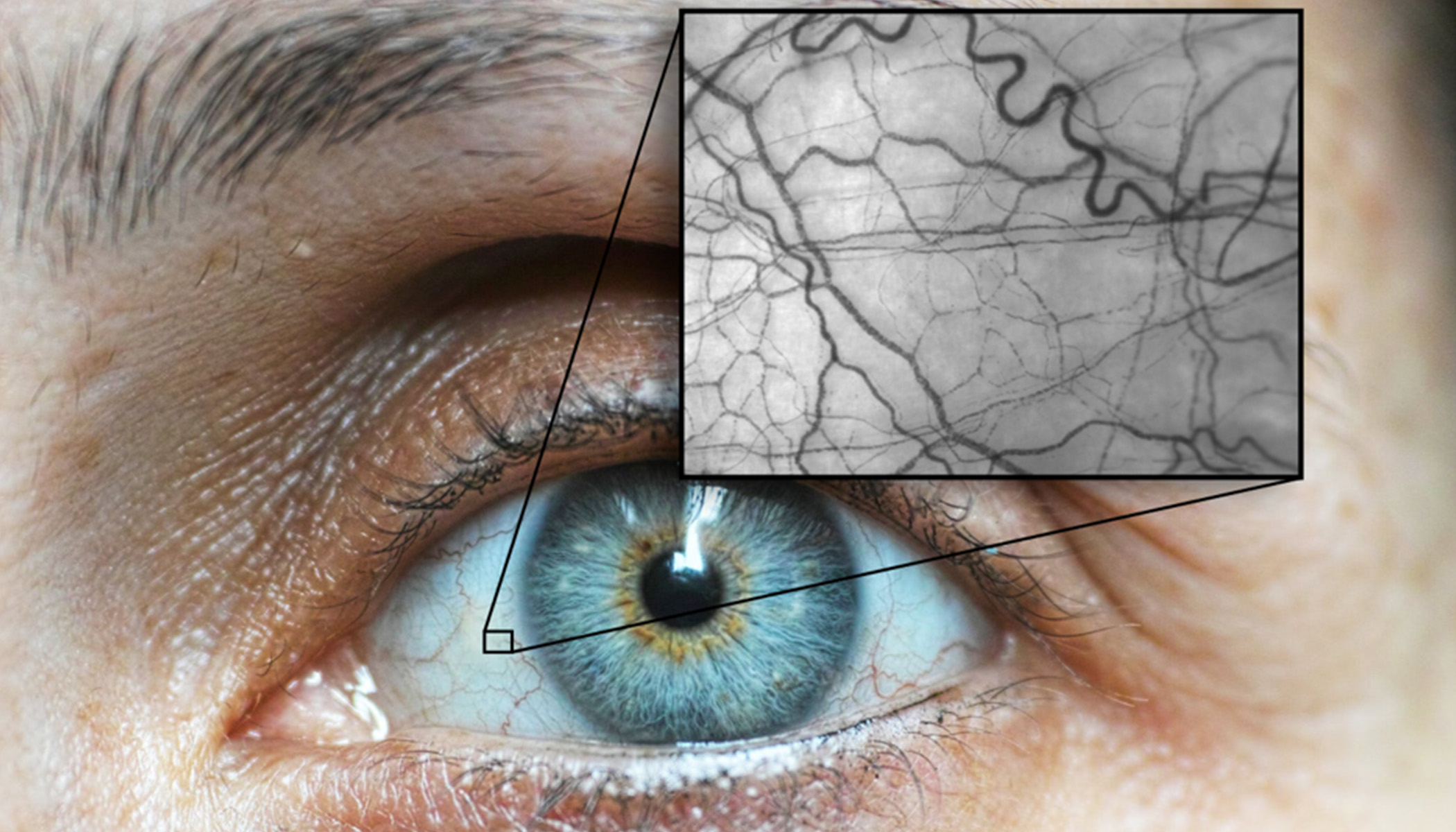
This autofocus technology has been used to study ocular microvascular blood flow with single cell resolution. It should also be applicable to the analysis of any living organisms and other moving microscopic objects.
Opportunity
We wish to license the technology.
Inventors
Paul Meyer MD FRCP
Retired Consultant Medical Ophthalmologist, visitor in the Department of Engineering and Senior Visiting Fellow in the Department of Medicine, Cambridge University.
In the 1980s, Prof. Paul Meyer started the Addenbrookes Hospital Medical Ophthalmology Unit, where he remained a consultant until his retirement in 2017. His interests included inflammatory eye diseases and the relationship between ophthalmic pathology and systemic disease, particularly systemic vasculitis, and the evaluation and management of thyroid eye disease.
During his MD, Prof Meyer demonstrated immune complex formation and deposition in ocular surface microcirculations by image-intensified videomicroscopy and characterised patterns of blood flow in the anterior segment of the human eye. He developed 3 new angiographic techniques, including haemoglobin video imaging (HVI), which has since been used to evaluate systemic disorders and the effects of systemic therapies on microcirculations. More recently, it has been adopted in glaucoma research to provide a surrogate for the quantification of aqueous outflow.
His current research focuses on the use of the ocular surface vasculature as a sample microcirculation to establish systemic and ocular diagnoses and to evaluate responses to treatment in systemic metabolic and inflammatory diseases.
Outside ophthalmology, Prof. Meyer has invented a variable abduction hip brace, now widely used in the management of cerebral palsy. He also developed a novel seating system and wheelchair, a gravitational sitting-to-standing frame and various other devices for physically disabled children.
Dr Thierry Savin, Associate Professor in the biomechanics group at the Department of Engineering
Within bioengineering, Dr Savin focuses on developing theoretical and experimental methods to study complex fluids and living matter over a wide range of scales. The goal of this work is to uncover the physical rules explaining the macroscopic properties and functions of materials, tissues and organs from the behaviour of their ‘elementary’ constituents.
Dr Savin’s research interests include:
- Structure, mechanics and thermodynamics of living matter
- Biophysics
- Materials Science
- Transport Phenomena
Thomas Lynch, Research student in the biomechanics group at the Department of Engineering