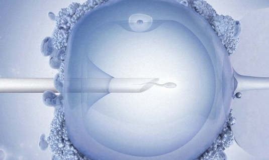
Researchers at the University of Cambridge have developed a new technique which could significantly increase success rates of pregnancies and reduce the frequency of multiple pregnancies associated with in vitro fertilisation (IVF).
The research has found that a series of movements in the egg in the first hours after fertilisation can be used to predict which embryos have the best chance of survival. The findings were published Tuesday (9 August) in the journal Nature Communications.
Normally, embryos are implanted after two or three days in culture, with their progress at this stage used as a predictor of their likely development. However, around half of all human embryos stop developing before the blastocyst stage at day five of development. As a result, patients are often implanted with many embryos at once, which can lead to multiple pregnancies.
It is important to be able to quantitate some indication of the health of embryos at the earliest possible stage in order to minimise the time they have to spend outside the body of the mother.
Professor Magdalena Zernicka-Goetz
In order to improve success rates and reduce the number of multiple pregnancies, which are potentially risky for the mother and often end in miscarriage, many IVF clinics delay implantation until embryos have reached the blastocyst stage, and implant fewer embryos per round of treatment.
“There is a need to ensure that the embryos which are implanted into the mothers are those which stand the greatest chance of resulting in a successful live birth,” said lead author Professor Magdalena Zernicka-Goetz of The Wellcome Trust/Cancer Research UK Gurdon Institute and the Department of Physiology, Development and Neuroscience at Cambridge University.
“It is important to be able to quantitate some indication of the health of embryos at the earliest possible stage in order to minimise the time they have to spend outside the body of the mother.”
Professor Zernicka-Goetz with her team and her co-authors from Oxford and Cardiff Universities have developed a non-invasive method of accurately predicting the likelihood that an embryo will develop not only to the blastocyst stage, but also to birth from as early as two hours after fertilisation. “This outcome represents years of work in building the necessary experience and bringing a team of talented scientists together, but it has been well worth it,” she said. “When we obtained the first promising results nearly five years ago, we applied for funding from the Wellcome Trust and fortunately were successful and so could continue our work.”
The researchers discovered that fertilisation initiates a series of pulsating movements in the cytoplasm, the jelly-like substance which fills the cell. The movements are caused by an interaction between the sperm and the cortex of the egg, and coincide with pulsations of the fertilisation cone, which is formed when the sperm and egg plasma membranes fuse to draw the sperm into the egg. By constructing time-lapse recordings of the cytoplasmic movements and pulsations of the fertilisation cone over a period of two hours after fertilisation, using differential interface (DIC) microscopy and using a sophisticated image analysis technique called particle image velocitometry (PIV), the researchers charted the frequency of these movements in a study involving mouse zygotes. These recordings showed a linear relationship between the speed of the movements and the likelihood that an embryo will develop to the blastocyst stage. This allowed the group to develop a mathematical model enabling them to predict successful development beyond this stage – all the way to birth.
Improving success rates for IVF by choosing the best embryo to implant not only decreases the risks associated with multiple embryo transfers but also potentially minimises the number of rounds of IVF that potential mothers must go through in order to achieve a successful pregnancy.
Dr Steven Suchting
Since similar movements occur in human embryos, these movements should be similarly predictive of embryo viability in humans. The invention provides a quantitative method of determining embryo viability which is much more reliable than current qualitative visual inspection. The inventors are being supported in their commercialisation efforts by Cambridge Enterprise, the University’s commercialisation group.
“In vitro fertilisation is an emotionally and physically demanding experience for prospective parents, with current success rates of only 30%,” said Dr Steven Suchting of Cambridge Enterprise. “Improving success rates for IVF by choosing the best embryo to implant not only decreases the risks associated with multiple embryo transfers but also potentially minimises the number of rounds of IVF that potential mothers must go through in order to achieve a successful pregnancy. This saves prospective parents both the trauma and cost of the IVF experiences.”