
Both the ET and MF murine models have been robustly characterised, and are valuable preclinical models available for non-exclusive licensing.
Paper
Mutant calreticulin knock-in mice develop thrombocytosis and myelofibrosis without a stem cell self-renewal advantage, Green et al., Blood, 2018, doi: 10.1182/blood-2017-09-806356.
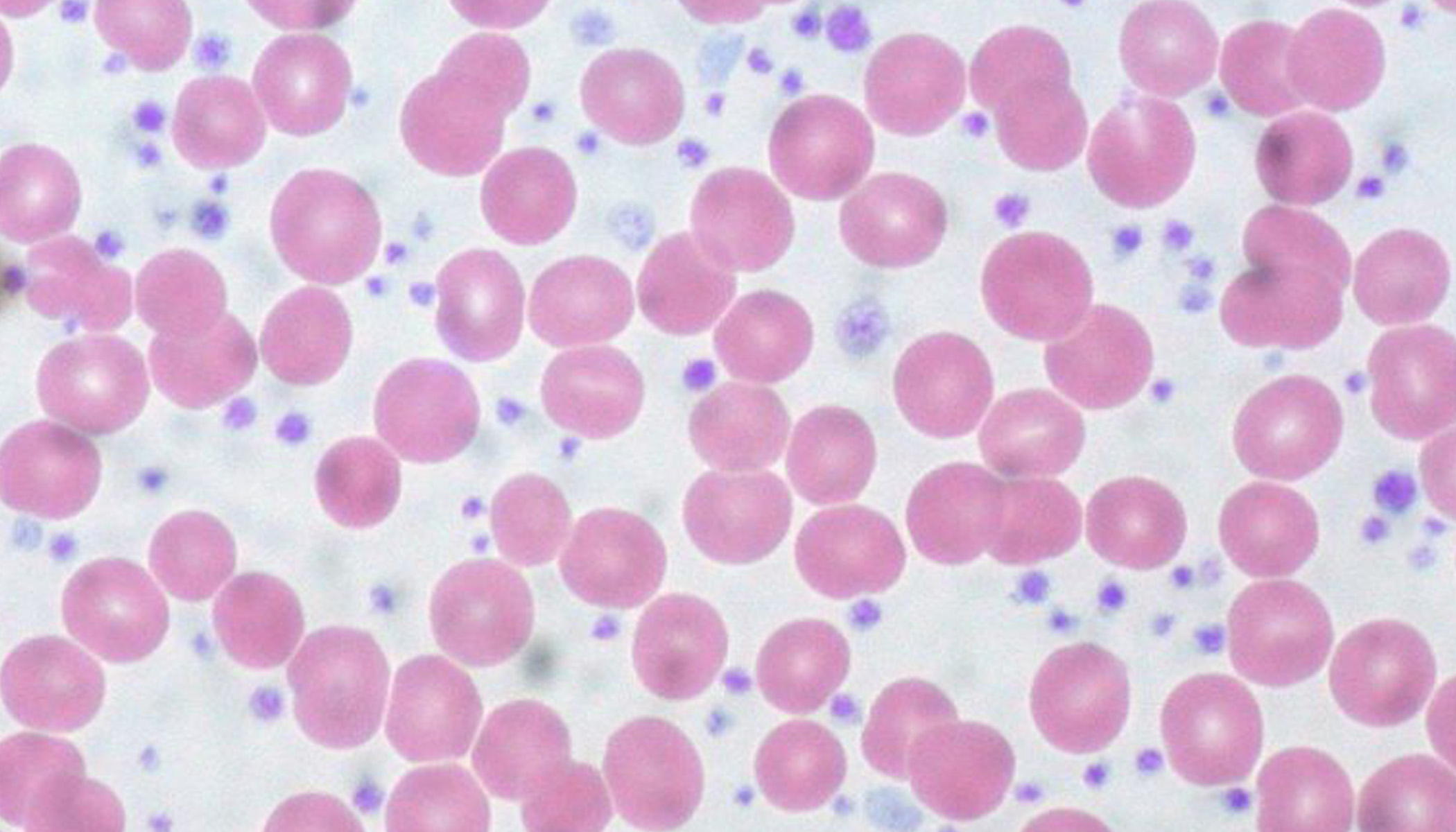
Banner image: Essential Thrombocythemia, Peripheral Blood, from Ed Uthman via Flickr, Attribution (CC BY 2.0)
Mouse Models of Essential Thrombocythemia and Myelofibrosis - Available for Preclinical Studies
Researchers at the University of Cambridge have generated fully characterised, conditional knock-in mouse models for essential thrombocythemia (ET) and related myelofibrosis (PMF) that are available to license for preclinical studies.
This mouse line conditionally expresses a mutant Calreticulin with humanized novel C terminus which is found in human patients, under the control of the endogenous mouse locus.
Technology overview
Essential thrombocythemia (ET) and Primary myelofibrosis (MF) are two types of myeloid proliferative neoplasms (MPNs). MPNs are chronic hematologic malignancies that arise in the hematopoietic stem cell compartment and share a variable propensity to transform to acute myeloid leukaemia.
In ET and PMF, 50-60% of patients carry a JAK2 mutation. Of the non-JAK2 mutated patients, 70-80% carry mutations in calreticulin (CALR) which encodes an endoplasmic reticulum chaperone.
Key Features
- Mutant CALR expression results in expansion of immunophenotypic HSCs
- Shows clear differences in the consequences of CALR and JAK2 mutations
- Powerful tool to further dissect mutant CALR contribution to myeloid proliferative neoplasm (MPN) pathogenesis
- Enables screening and preclinical validating of drugs targeting MPN stem cells
ET Model
- Heterozygous CALRdel/+ C57BL/6
- Develops a myeloproliferative phenotype resembling human ET
- Presents with marked thrombocytosis
- Displays increased megakaryopoiesis
PMF Model
- Homozygous CALRdel/del C57BL/6
- Presents with severe thrombocytosis and development of MF
- Presents with an increased white blood cell count and reduced haemoglobin level
- Presents with splenomegaly
- Displays increased BM reticulin level